Week 2:
University of Chicago Medical Center
July 2, 2013
Fellow in Research
Monday of week two was great! First off we worked on de-fatting the cerebellum slides. Being as fat is hydrophobic, it gets in the way of the luxol fast blue staining the myelin. We placed the slides in the luxol fast blue dye overnight. Then I got to watch Aya and Kae harvest bone marrow cells! The RPMI media I made last week was actually used to harvest the cells! We only want the bone marrow cells so to get rid of the red blood cells we added red blood cell lysis buffer to cause the red blood cells to burst. After watching it a few times I even got to add lysis buffer and centrifuge the cells myself.
After we removed as many of the red blood cells as we could, we removed a sample of the bone marrow cells and looked at them under a microscope. I actually got to count the bone marrow cells so we could get an estimate of how many cells we had harvested. Made me remember my cell culture dishes from BIO 141! We harvested approximately 240 million bone marrow cells!
Tuesday I got to continue with the cerebellum slides. I prepared one of the solutions for the luxol fast blue stain. Luxol fast blue is a staining technique that hasn’t been used much in this lab so I got to kind of experiment with it to figure out what would produce the best results. This is what the finished product looked like.
Aya was working on cell culturing so I got to observe that first hand! For that she was primarily concerned with the hippocampus, which was sliced super thin and placed in wells. This allows us to have cells that can be manipulated as need be and after a few weeks these cells are basically the same as cells in an adult rat.
Kae and I then worked with Q-dots, which basically are these tiny nanoparticles that can bind to other particles of interest and can fluoresce lots of different colors!
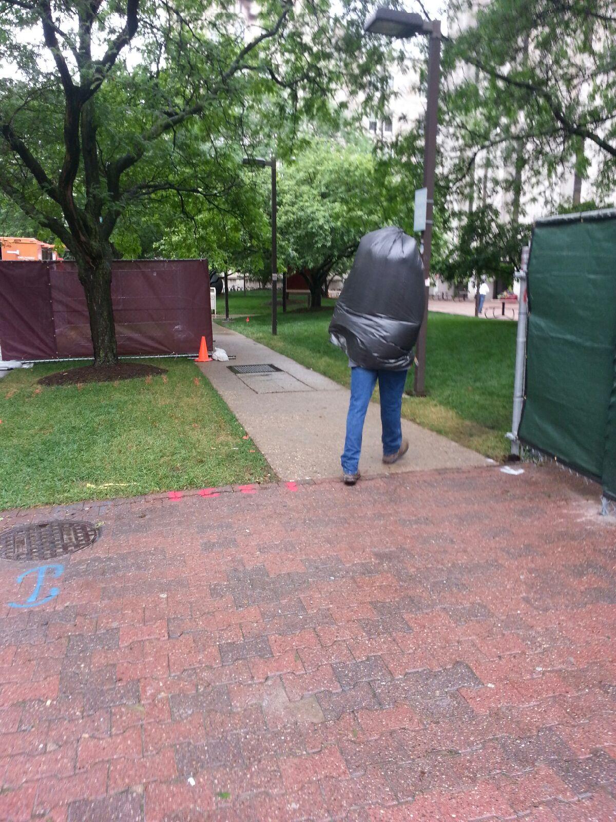
Wednesday on the way to work I saw this person in the rain.
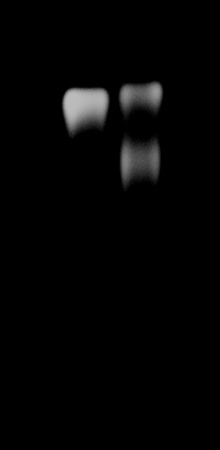
Once in the lab, I got to learn how to make electrophoresis gels to run antibodies on. I loaded in a control Q-dot sample and the Q-dot sample Kae and I made on Tuesday which we conjugated to an antibody called CD63. This is the gel, the separated band tells me that the Q-dots conjugated to the antibodies did bind because the control on the left containing unconjugated Q-dots only had one band and the Q-dots conjugated with antibodies had two bands. The top band is the Q-dots that did not conjugate to CD63 and the bottom band is the population of Q-dots that did bind to CD63.
I also got to learn how to do a BCA protein assay, which I found out will be pretty important to my project! I had samples of unknown protein concentrations so I had to use a set of standards to then be able to compare the unknowns to, which that part was similar in concept to a couple of my analytical chemistry labs at Cornell.
Thursday I ran another gel but this time we cut out the part of the gel that contained the extra band. This band is our conjugated antibodies and Q-dots. We used a special filter to extract conjugated antibodies and Q-dots from the gel. Now we have our pure population of Q-dot-CD63 ready to attach to our exosomes!
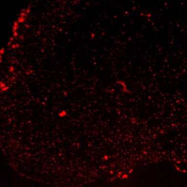
Friday we used the conjugated antibodies and Q-dots to tag the exosomes. Once we tagged the exosomes, we introduced the tagged exosomes to the slice cultures. The Q-dots fluoresce, so when we looked at the slice cultures under the microscope we could see where all of the antibodies traveled by the red dots! The red dots inside the cell mean that our tagged exosomes were taken in by the cells in the culture slices.
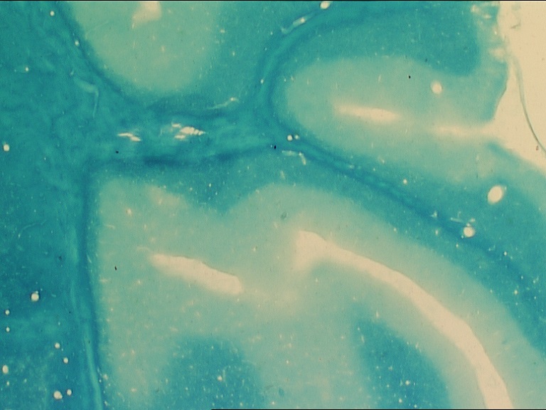
Then I got to look at the luxol fast blue slides I had been working on all week under a microscope! The dark blue areas are white matter and the light blue areas are grey matter.
Most of the other students in my program have either already taken the MCATs or are currently in the process of studying for the MCATs. They have been super great about offering me advice concerning my own preparation for my MCATs as well as sharing some of their practice materials. All of the other students in the PSOMER program are either going to be a senior this upcoming year or are working towards their masters. It is nice having another year to prepare my medical school application and I will be able to ask any of the PSOMER students for advice.
This week I got a lot of hands-on learning. Seeing how everything is done and why certain things are done has helped me get a better grasp on our project. It’s strange to think that I’ve already been here two weeks already! I keep learning new things everyday, which has been awesome!!
Major: Biochemistry & Molecular Biology and Psychology. Hometown:DeWitt, Iowa.
PreviousWeek 1: University of Chicago Medical Center
NextWeek 3: University of Chicago Medical Center