This past week has been quite an adventure, with every day being both informative and entertaining. Here in the lab, there is no shortage of laughs between our projects, and sometimes even during them. Everyone here is incredibly friendly and funny, and they really make home feel like it's not almost 800 miles away.
My weekly saga began on Sunday, June 9th, with Cameron Reimers and I trying to hike to the Devil's Head Lookout tower. This trail is 1.4 miles long (one way) and about an 870 foot elevation gain. I was so excited for it, since we don’t really have many elevation gains like that in Iowa. Cameron and I got ready to head out at about 10:30. However, before we went hiking, we obviously had to get some good food in us; so we stopped by “Sams #3” in downtown Denver, right by Cameron's apartment. It was apparently featured in the hit television series, ‘Diners, Drive-ins, and Dives’, and we could see why. I ordered the french toast and was absolutely blown away by the taste.
After we finished there, we headed out to the trail. There was a chance of thunderstorms, but as we’re quickly finding out here, the weather changes every 5 minutes. Therefore, we really didn’t pay it any heed. In fact, when we got there, it was beautiful out.
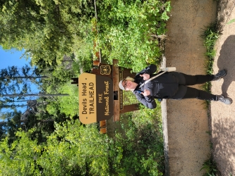
Me and the beautiful weather out at Devil’s Head Lookout.
We hiked for a bit over an hour and a half, and Cameron was able to make it to the top right before the tower closed due to the anticipated storm. I was taking it rather slowly due to the altitude, and taking plenty of pictures along the way. I was about 0.3 miles out when Cameron doubled back, letting me know that it was now closed. That’s when we both headed back down.
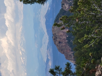
The view from a rock I sat on for a while
During the next few days at the lab, we made some acrylamide gels to fill up our supply in Dr. Wlodarchak’s lab. Chase, Dr. Wlodarchak’s research assistant, was able to help us some, but that didn’t stop us from doing the wrong step once or twice. Let’s just say that we got really good at doing the dishes that day. We also learned about protein purification, and we were able to assist Dr. Wlodarchak and Chase in their attempt. Unfortunately, as we found out later by running a gel, somewhere in the process we seemingly discarded the protein. We were again given the opportunity to split some cells; something I find I’m getting very good at.
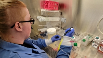
Splitting cells for the second time
I was able to spend some more time with Melissa, Dr. Keller’s lab assistant, on Wednesday; she stayed late to prepare cells for a flow cytometry demonstration for me and Cameron. She spoke a lot about the work they were doing with rats and about how the current results were a little surprising, as they found unexpected correlations between impacted organs. I was also able to witness cell staining of the spleen and PVAT (perivascular adipose tissue).
While all of this has been very fun and enlightening, there were two high points for me this week. First off, I found out what my research project for the summer is! I will be with Dr. Wlodarchak attempting to clear the water-bound tuberculosis strain M. marinum from a set of macrophages without destroying all the cells. I will do this by first growing the M. Marinum with 3 different fluorescent tags; mWasbi, tdTomato, and cerulean. This will take about 11-12 days of care and will result in being able to test which tag will show up best in our KEYENCE immunofluorescent microscope. This means that we will be able to see when all of it is gone after I attempt a clearance assay. I just started growing it on Friday, so stay tuned for the rest of the steps and to see how it goes!
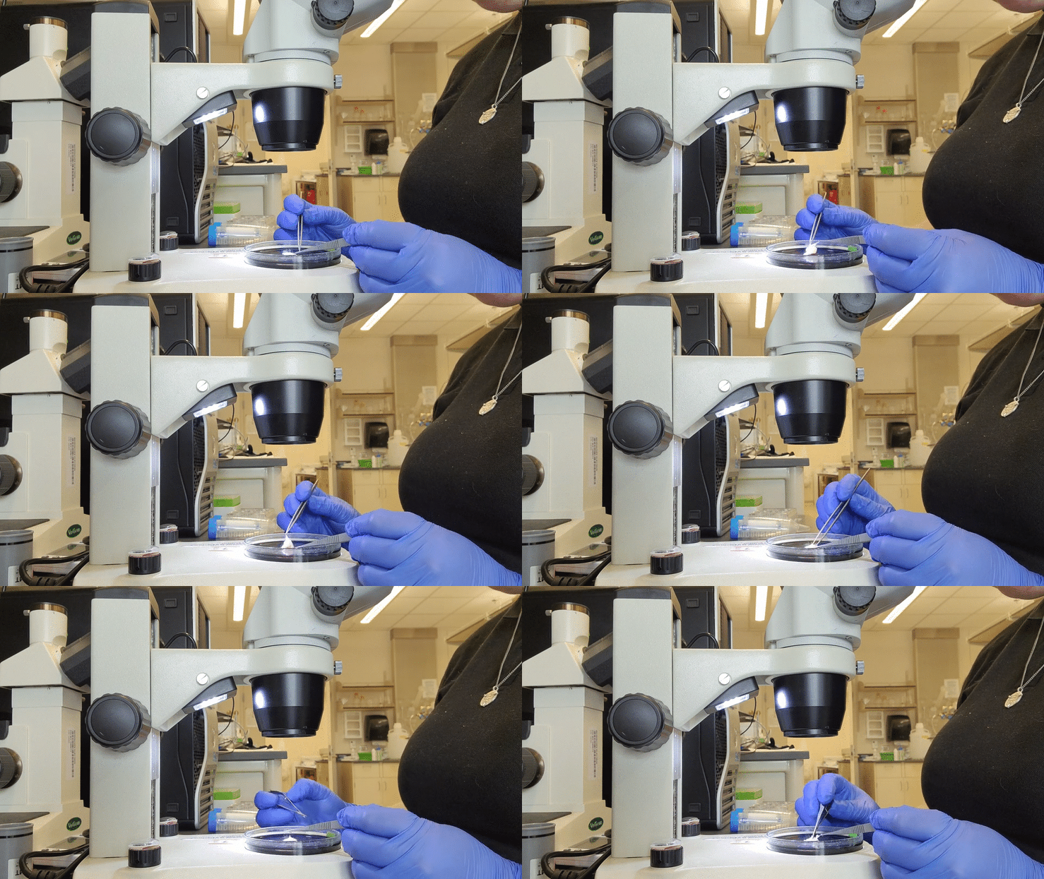
The second high point was when Dr. Keller stopped by to ask Cameron, Kevin (Another student intern), and I if we wanted to attempt a rat femoral aorta dissection, as she had some extra ones. We all jumped on it and got to work. She explained that she was doing this for a myograph, if we would like to give that a shot as well. We began by dissecting a clump of fat smaller than the tip of my finger to begin looking for the femoral aorta. She promised it was in there, but the first 10 minutes we could have sworn she was lying to us. Regardless, she had obviously been right, and we were able to find it after a bit of careful digging.

Cameron took a photo of me engrossed in the work
From there and despite it being at the end of the day, Dr. Keller let us attempt to put the aorta on the myograph cannula. It took a few minutes, but we were able to do it.
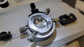
A close up of a rat aorta, half attached to the myograph machine
The next day, she left us with a few more of the same samples to continue practicing on if we had wanted to. I was incredibly excited to try my hand at it again, now that I knew what I was doing. It only took me 25 minutes this time to do it, which is so far a personal record. I’m sure it helped that this piece had been bigger than the last.
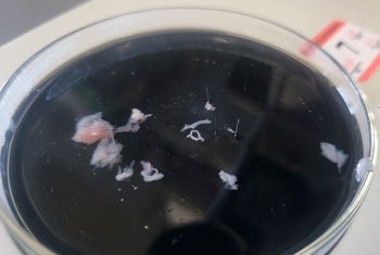
The middle top is the femoral aorta, and the middle bottom is the saphenous nerve; the rest is fat and membrane
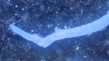
A close up through the microscope lens of the femoral aorta in a rat
All in all, I have learned so much this week! From an introduction to flow cytometry, to dissections, and all the way to designing my own experiment, this week has been full of adventure! I can’t want to see what next week brings, and I can’t wait for my bacteria to finish growing! See you all next week!