Week 2:
Training
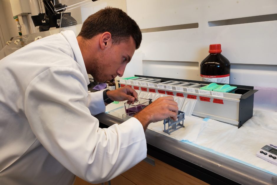
Paul using a staining process know as Cresyl violet staining or Nissl staining to expose Nissl bodies on tissue slides cut on Cryostat
June 9, 2019
In addition to completing Compliance Training last week, I spent a lot of hours of my day reviewing neuroscience material to get myself familiar with terms and mechanisms in neuroanatomy that are necessary for understanding the project I am observing as well as my own project. I also have been reading a lot of scholarly articles that talk about specific pathways in the nervous system focusing on nociceptive and neuropathic pathways, which is helping me understand my project. After a week of getting myself familiar with the scholarly material, I met with Dr. Stephen Onifer, PhD, the head scientist of the science research project I am observing. We went over exactly what my project is going to be and how I am going to carry it out. We established that during my project I am going to learn about 1) the mobility of connective tissues in the lower back known as thoracolumbar fascia and 2) sensory neurons that transmit information from the thoracolumbar fascia to the spinal cord. I then attended a meeting with clinician scientists about research being planned to study the mobility of the thoracolumbar fascia in chronic low back pain. I was also introduced to the clinicians and toured the clinical research facility and saw all their equipment that will be used in this research study.
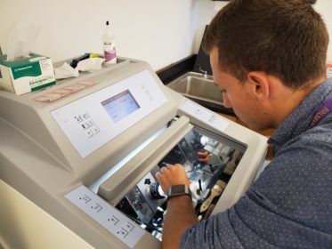
This week I also performed a lot more hands-on procedures than just simply watching procedures being done. Like last week, I watched some of the scientists embed tissues into molds for freezing and subsequent processing. After some observation, I was able to embed my own tissues into molds. Even though my first embedding procedure was a little rough, I was able to learn and got a lot better at it in the following days when I did more embedding. Then one of the scientists taught me how to use a machine called a cryostat which cuts tissues embedded in the frozen molds into very thin sheets so that they can be placed on microscope slides for staining and viewing with a microscope. Similar to the tissue embedding, my first practice run with the cryostat did not go great, but again, I was able to learn and in the subsequent days I ended up getting a lot better at tissue placement along with my operation of the cryostat. After being taught those two skills, one of the scientists commented on how impressed he was with my ability to learn things quickly. I think Cornell’s accelerated curriculum and liberal arts academic process, where I have to sometimes adapt to complex problems on the fly, has really helped in developing my ability to learn new things quickly and effectively.
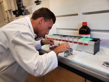
Along with researching, observing, and learning new skills, I conducted an informal interview with Dr. Robert Vining, DC, DHSc, the Associate Dean of Clinical Research at the PCCR. I was able to gain insight on his career path, which includes private practice, teaching, and research. I also got some insightful information to help me think about choosing a chiropractic college and different ways to think about chiropractic techniques. Lastly, after all of the processes of embedding tissues and then cutting them on the cryostat was over for the week, it was time to start staining tissues. I used a process called cresyl violet or nissl staining that allows me to see nissl bodies in cells within the tissues. With the help and teachings from one of the chiropractic students working on her project for the R15 Research Honors Program, we performed the staining procedure with tissues on some of my microscope slides. Next week when the slides have dried, we will look at the stained tissues with a microscope and see the results.
Paul is a kinesiology major with a minor in biology from Leavenworth, Indiana.