Week 3:
Learning New Skills
June 16, 2019
This week, I was very busy with a lot of different tasks, such as cutting tissues on the Cryostat and placing the sections on microscope slides, staining the sections, inspecting stained and unstained sections with a microscope, meeting with my basic science mentor, Stephen Onifer, PhD, reading scientific articles, and applying to Chiropractic colleges. Throughout the week, I set up the Cryostat with the help of a scientist Mr. Randall Sozio, B.S. This involved removing blocks of frozen tissues in plastic molds from an ultra-low freezer and attaching each block to a metal holder. I then observed Mr. Sozio cut the blocks with the tissues. After he was done cutting his sections, I would hop on the Cryostat and start cutting my own sections.
At the beginning of the week my sections placement was a little sloppy. By the end of the week, I was able to become more precise and faster with my placement, and my slides turned out very good. I am glad that I have started to get the hang of this new skill. I then spent some time inspecting both stained and unstained sections on microscope slides. While cutting, I would periodically look at the sections with a microscope, comparing what I saw to images in an atlas. With the help of Mr. Sozio, I got a sense of where I was in the tissue to get exactly the right sections needed for my project. When we were done with the Cryostat, I would take my slides and put them in the ultra-low freezer. Then I would help Mr. Sozio clean up the machine and the lab area before we locked up for the day. After my cresyl violet-stained sections from last week dried, I inspected them as well. The staining appeared faint but after adjusting the contrast of the microscope they were easy to see. They ended up turning out pretty good.
This week, I also started meeting with Dr. Onifer about every morning, where he and I would go over my presentation guidelines and PowerPoint presentation. He is helping me to develop the presentation, making edits and adding in suggestions that we think will make the it turn out just right. My time out of the lab was spent with some scientific articles Dr. Onifer had given me. These will go in conjunction with a lab gathering called a Journal Club that Dr. Onifer will set up next month for Palmer chiropractic students in the R15 Research Honors Program and faculty. There, we will talk about scientific articles in a group setting. For this Journal Club, I will be leading the discussion. At first, I will teach about a neuropeptide, calcitonin gene-related neuropeptide (CGRP). Then, I will talk about a scientific article involving CGRP summarizing it for everyone. I am nervous to lead this discussion but with Dr. Onifer’s guidance and the group discussion skills I have obtained from classes at Cornell I know I cannot fail.
Later in the week, Dr. Onifer treated me and the laboratory to a meal at Buffalo Wild Wings. It was a fun time with good people and good conversation. During that outing they also showed me around different parts of Davenport, IA, since I have never really been in this area of the city which was a good time on its own. To round out the week, myself and another scientist Mr. Charles Ahrends, B.S., took microscope slides with sections that I had cut out of the ultra-lowfreezer and brought them to the tissue staining lab.
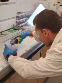
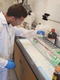
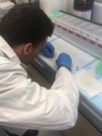
In the lab, Mr. Ahrends, accompanied by Mr. Sozio, had me set up the whole cresyl violet staining process by filling up all the containers with their respective chemicals and getting the slides ready to be stained. It was a daunting task at first but I managed to complete it and did everything by the protocol and as I had remembered from staining last week. I also made sure I was doing everything correctly by asking Mrs. Ahrends and Sozio when I had questions or was not sure about a step. After everything was set up, I continued the staining process on my own with the helpful oversight of the two scientists by putting the slides in each chemical container for a set amount of time.
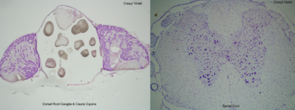
Once I finished the staining process, I put coverslips on each slide and sat them in a slide book under the chemical hood to dry. When that was finished, Mr. Ahrends took into the microscopy room where a microscope is hooked up to a computer so that we can take pictures of the sections on the slides. We took pictures from the slides I stained last week. Then, we constructed a montage of pictures you can see below.
Along with reading articles during my time out of the lab, I have been contacting admissions offices at different Chiropractic colleges, such as Palmer, here in Davenport and in Florida, and Life University in Georgia. I also started the application process for Palmer in Davenport From an already quick transcript evaluation from their admissions team, they already like what they see. They said if I start my application process now, I could be accepted before my senior year at Cornell even starts, which is great news. I have the support from everyone around me and Cornell College to thank for it. It turned out to be a pretty eventful week. I cannot wait to get started on next week’s projects.
Paul is a kinesiology major with a minor in biology from Leavenworth, Indiana.