Week 4:
Mastering a Skill and Making Connections
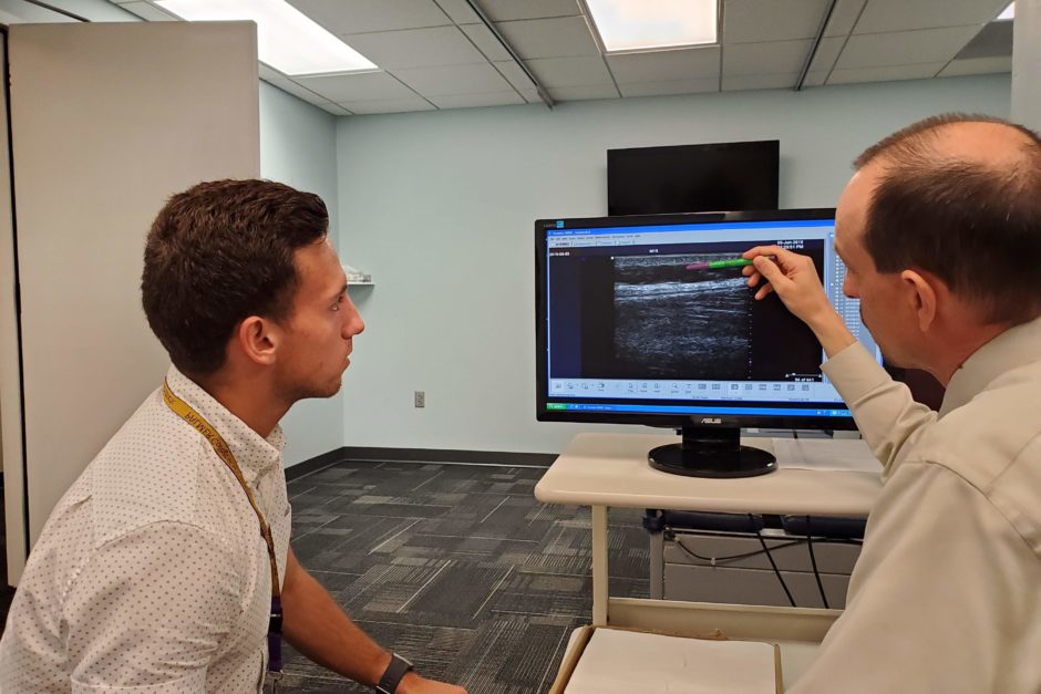
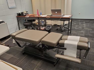
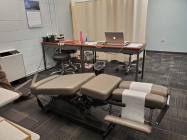
Dr. Vining explaining the different levels of tissue in the lower back that are exposed with the ultra sound device during their clinical study.
June 23, 2019
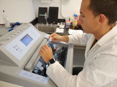
This week, I mainly performed one of the skills that I learned during my time here at the PCCR, cryosectioning. Additionally, I went from “bench to bedside” to Palmer’s Chiropractic Research Clinic. There, I met some of the Clinician Scientists and was able to learn first-hand about a method they are developing. Every day this week, except for Friday, was mainly filled by me setting up and cutting tissues with the Cryostat. Unlike last week when I cut spinal cords, this week I cut a new tissue that I had never done before, dorsal root ganglia or DRG. These contain sensory nerve cells (neurons) with fibers (axons) in roots that terminate to communicate with neurons in the dorsal region (horn) of the spinal cord and with axons in spinal nerves that then branch out to form sensory nerves in your peripheral nervous system. For this cryosectioning, I used frozen DRGs that I previously helped to prepare.
Compared to the spinal cords, the DRGs were a little harder to get used to. The sections are wider, requiring a different placement on microscope slides. Once I got the hang of placement, my precision improved and cutting went a lot smoother and more quickly. I think that by the end of the week, I managed to cut DRGs to fill up around one hundred and thirty slides. When I finished cutting all the DRGs, we moved on to another round of cresyl violet staining.
Unlike last week, Mr. Sozio set the staining station up for me. I ran some of the slides through each staining step and cover-slipped them all on my own. After staining was finished and cover slips were put on each slide, I left them to dry over the weekend under the chemical hood so that we can look at them with the microscope next week to prepare for immunostaining sections that I cut on other slides to see CGRP. The ccryosectioning and staining procedures are being done so that we can get a more specific and magnified look into the DRGs, using a microscope. The neurological mechanisms by which chiropractic produces analgesia are largely unknown. One set of techniques available to chiropractic researchers investigating potential neurobiological mechanisms is histology and microscopy.
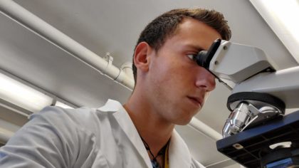
Examining tissues at a macroscopic level provides general information. Peripheral nerves, spinal nerves, DRGs, roots, and the spinal cord to which they are connected can be seen without a microscope. However, anatomy at the cellular level and visualizing some aspects of nerve requires a microscope and specialized tissue staining techniques. Cutting DRGs and staining the sections with cresyl violet allows me to see cells and organelles, such as their nuclei, in them with a microscope. Visualizing organelles in more detail as well as cell types (neurons, glia), parts (cell bodies, processes), structures (filaments, matrices), and the chemicals cells make to function and communicate with other cells or tissues (enzymes, neurotransmitters) can be done by immunostaining the sections. For example, I am learning about calcitonin gene-related peptide (CGRP), a neurotransmitter made by DRG neurons that may play a role in spinal manipulative therapy analgesia.
I will use the microscope to look at the sections stained with cresyl violet. After finding microscope slides that have DRG sections, I will immunostain other slides with adjacent sections to see neurons that made CGRP. Using histology and microscopy techniques like these along with special analyses procedures allows chiropractic researchers to address questions such as “Do the number of cells making neurotransmitters like CGRP change following spinal manipulative therapy?”. If the number of cells change, then we come closer to understanding some potentially important neurological mechanisms and further hypotheses can be generated and tested.
Through my research, I am learning scientific processes and methods that will inevitably help in other research settings that I will be put in at chiropractic school. This internship is giving me an opportunity to get a more in-depth view of anatomy and physiology of the peripheral and central nervous systems. When I do become a chiropractic student and then practitioner, this experience will he me better understand the biological mechanisms underlying manipulative treatments, helping me make more informed decisions about the care I will one day provide. The internship also will make me more competitive when I apply to chiropractic schools, since I will have a better understanding about some types of research that inform chiropractic practice.
I also got the chance this week to walk over to Palmer’s Chiropractic Clinic and talk with some of the chiropractors. I mainly spoke with Dr. David Juehring, and Dr. D. Ranier Pavlicek. Dr. Juehring oversees the Rehabilitation and Sports Injury Department. It was a very enriching experience to talk to other chiropractic practitioners besides those at the PCCR and in my family, to see what they do and how they practice, and to make some new connections. Being an athlete all my life, I really want to work with athletes when I become a chiropractor and I want to develop an environment that incorporates sports medicine and is also an area that can help educate athletes on the importance and possible benefits they can receive by seeing a chiropractor. So, during our conversation I was able to talk with them about my ambitions of how I would like to practice. They were able to give me some helpful advice on how to start that path, such as certain certifications I would need. They also gave me the contact information of two chiropractors that are practicing in a very similar way to how I want to so that I could get some insight on what it is like to practice in that certain way. Overall, the interaction was a great experience and I am glad that I have made some new connections with people in this field.
I also had the privilege this week of going down to the clinical research division of the PCCR to be involved in the study that Dr. Vining and other members of his team are doing, but as a participant. For this study they are looking at the mobility of the thoracolumbar fascia connective tissue in the lower back when the body is flexed in a certain way using a special chiropractic table, known as a flexion distraction table, and they are looking at this connective tissue using an ultrasound device. Being a participant involved laying on a special chiropractic table, known as a flexion distraction table, where they would flex it up in down and then use an ultrasound device, placed on my lower back, to get an inside image of what was happening when they were flexing the table. It was a fun experience being a participant, and I am glad I could help out with their research and give them feedback on what they could do to help improve their procedure and during the experiment to help it go a little smoother. Next week, I will get the chance to observe this procedure happening instead of participating, so I am excited to see how that goes and I hope to get more clinical research exposure as the summer goes on.
Then to finish out the week, I had a meeting with my basic science research mentor, Dr. Onifer. I showed him the progress I had made with my PowerPoint presentation. He gave me some suggestions to consider for my presentation and we spoke about ways that I could talk through each slide when I present it. After the meeting, I typed up my blog post for the week and looked back over my notes on CGRP and the Journal club paper that I will be discussing during an upcoming lab meeting.
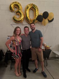
When I am not at the lab, you can usually find me spending my time at my Airbnb that I have rented for the summer. The Airbnb is in Milan, Illinois and I am living with my hosts who own the house. My hosts are young and welcoming people, and their house is wonderful and has basically every amenity that I could ever need. One of my host’s birthdays was this weekend, so his wife set a surprise party for him at this restaurant called the Armored Gardens, it was a really nice venue and the party was a blast. I met a lot of new people and made some amazing connections with some of my hosts’ friends. I am beyond thankful that I have meet such nice people through renting this Airbnb and I cannot wait to see what the rest of the summer holds. The rest of the weekend I finished up another training week for cross country with an eleven-mile-long run on Sunday and then spent the final moments of my weekend writing the rest of this week’s blog post and watching Avengers: End Game with my hosts.
Paul is a kinesiology major with a minor in biology from Leavenworth, Indiana.