Week 5:
All about Histology
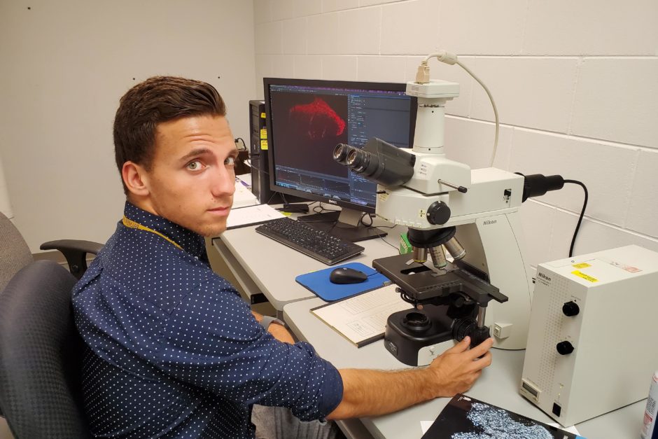
Me learning how to use the Microscope in the microscopy room. Section shown on computer in back ground.
June 30, 2019
Instead of tissue cryosectioning, this week’s histology theme was section staining focused. This included observing the stained sections with a microscope. The staining we performed this week is called immunostaining. Unlike cresyl violet staining where we expose cells in a general way, their shapes, and nissl bodies inside them, immunostaining is a technique used to penetrate inside cells and to get a deeper look at components within them. This is done by taking advantage of the antibody-antigen reaction, that is, when antibodies recognize their specific target antigens, they bind to them. In my case, the target antigen I am focusing on is calcitonin gene-related peptide (CGRP)., This neuropeptide is used by cells to communicate with other cells throughout the nervous system. Since we are looking at tissue sections with cells that make CGRP, immunostaining is a good method to see it.
The first couple days of this week, I observed Mr. Arends and Mr. Sozio fill out protocol sheets, mix chemical solutions, and pipet these solutions onto microscope slides to immunostain spinal cord sections on them. They were immunostaining for a protein (antigen) in spinal cord cells used to communicate a signal from the cell surface to the nucleus. A Palmer Chiropractic Student Research Investigator is studying this protein for his R15 Honors project. It was a good time to watch and learn before I did any hands-on work.
On Wednesday, we started my immunostaining procedure. I helped make up the chemical solutions using pipets to transfer stock solutions into other solutions, a vortex machine to mix them together, and a centrifuge to spin some stock solutions down to the bottom of their containers so that we could gather all of them in the pipets without having to worry about a portion of it being stuck on the top or sides of the container. For my first immunostaining step, I used a pipet filled with a buffer solution to wash the dorsal root ganglia (DRG) and spinal cord sections I previously cut and placed on the microscope slides. Then, I used blocking buffer which prevents the secondary antibody from binding to unwanted antigens. After adding the primary antibody to the sections, I left the slides alone for 24 hours so it had sufficient amount of time to bind to CGRP in the cells.
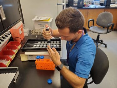
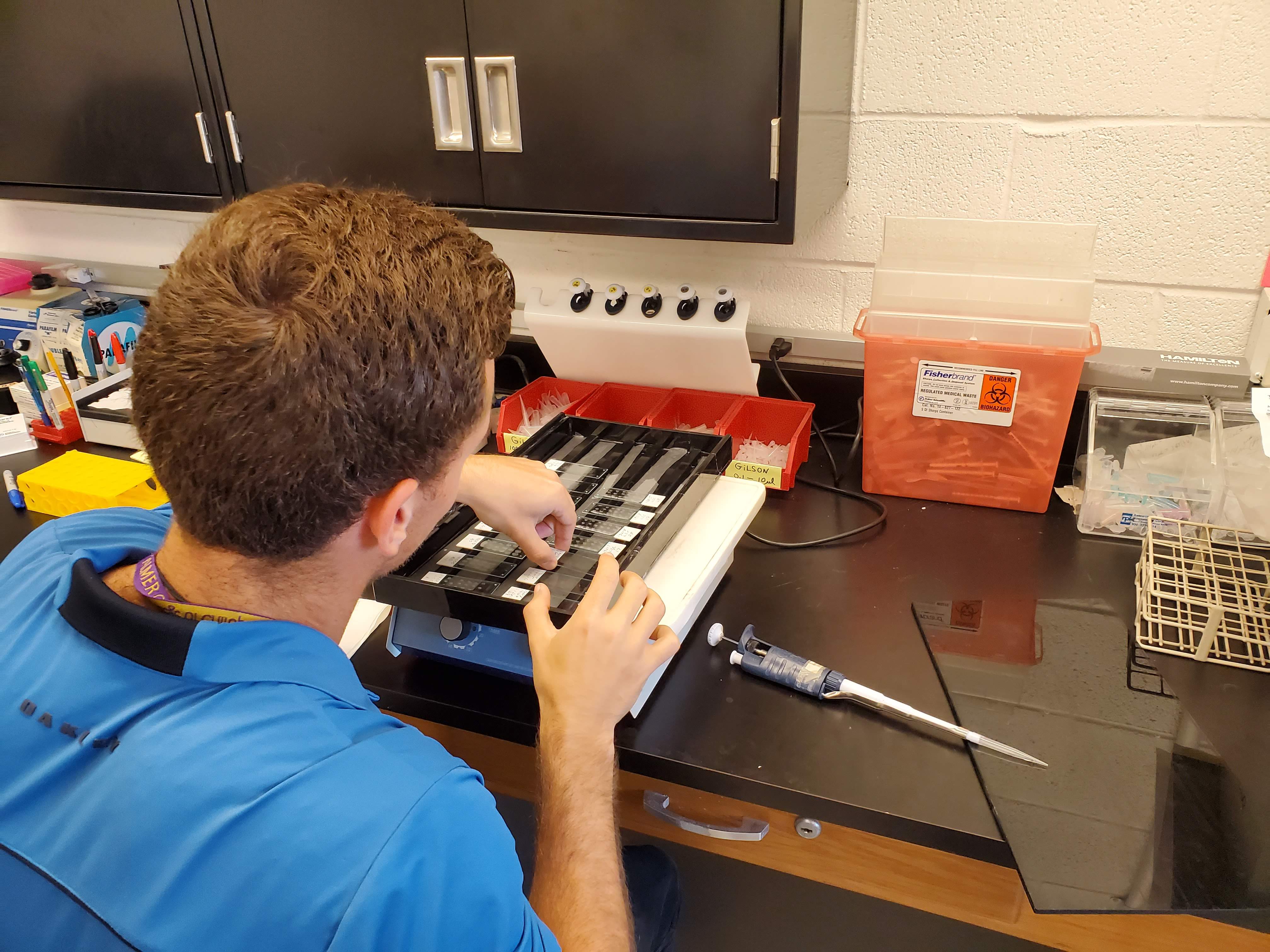
On Thursday, we started the second day of immunostaining by me preparing more chemical solutions, washing off the primary antibody, and then adding the secondary antibody so that it could bind to components of the primary antibody and allow me to see CGRP. I then washed the sections for the last time and placed coverslips over them.
On Friday, Mr. Arends and I went to the microscopy room where I observed the Palmer student use a microscope hooked up to a computer to takes pictures of his immunostained sections on slides. It was fun looking in the microscope to see cells in the sections in magnified detail and then on the computer monitor after a program was used to capture images of them.
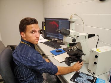
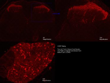
After the Palmer student was done, Mr. Arends and I first used the microscope to look at the sections we immunostained. To see CGRP, I shined light of a specific wavelength on the sections to excite a fluor attached to the secondary and primary antibodies. The immunostaing turned out extremely well. I saw CGRP in DRG neurons and their axons in the spinal cord. I took some good pictures and Mr. Ahrends and I made a montage to post to this week’s blog. It was exciting to finally see CGRP from tissues I embedded and in sections that I cut after talking about it for a month. This project has been fun and I cannot wait to continue it.
This week, I also had some meetings with my mentors, and had the privilege of going down to the research clinic and observe, while my basic science mentor, Dr. Onifer helped the clinical research team practice their ultrasound data collection technique. It was interesting to observe members of the clinical research team practice and develop their procedures. It was fascinating to watch the live imagery from the ultrasound device as it recorded the movement of multiple connective tissue layers in real time.
My first meeting of the week was with Dr. Onifer where he and I went over an outline and a PowerPoint presentation I am preparing for my Midterm and Final Assignments. I also had a meeting, later in the week, with Dr. Vining where we discussed improving the outline. Dr. Vining was also able to help me improve the flow of my PowerPoint presentation and challenged me to think about what was most important to communicate. The meetings were very constructive, and I think with the help of both Dr. Vining and Dr. Onifer I can vastly improve the quality of my presentation.
During my time outside meetings and the lab, I worked on my Assignments using the feedback of my mentors. I also looked at more scientific articles Dr. Onifer gave me. The articles discussed physical activity having an analgesic effect on chronic pain in the body, i.e. more activity, less pain. The articles were interesting, and the research is similar to what I would like to do someday. Also, this may be an area of study I will explore for my Kinesiology Research Honors project this year.
I also spent a lot of time with my girlfriend, Shay Rule, who came to visit me for the weekend. On Saturday, Shay and I went back toward Cornell College to visit a friend, Julian Smith, who is doing research with Cornell’s CSRI program. We met him at Coral Ridge Mall, and just hung out, got some food, and walked around the mall. After we got our fill of the mall, we got in Julian’s car and went to Iowa City to get some dinner, and then proceeded to walk around all of Iowa City, it was a really fun time and I hope we can all get together and do it again soon.
Sometimes I still can’t believe that I was given the opportunity to get this close to the chiropractic profession I love and get a behind-the-scenes look at the research that supports it. I am very thankful for this opportunity and I have my friends and family that support me, Cornell College, the donors that support the Cornell Fellows program, and the wonderful people at the PCCR to thank for it.
Paul is a kinesiology major with a minor in biology from Leavenworth, Indiana.
PreviousWeek 4: Mastering a Skill and Making Connections
NextWeek 6: Lab Time, Good Results, and a Well Deserved Break.